-
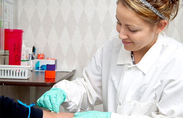
Phlebotomist Jennifer prepares patient’s arm to collect a sample.
-
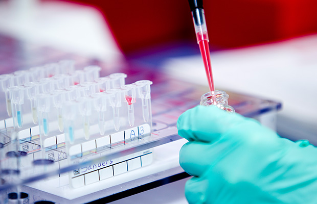
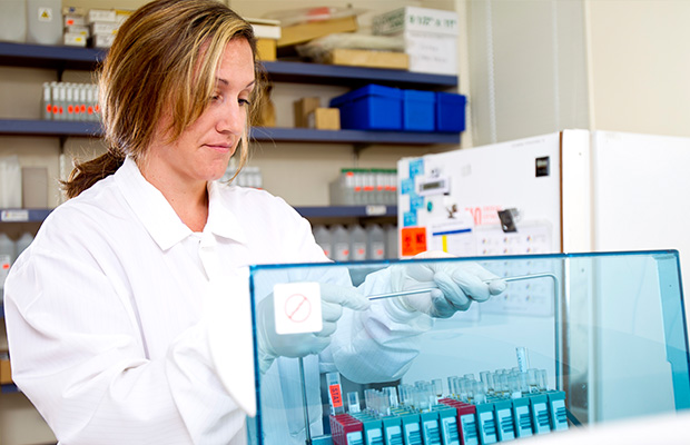
Technicians in the blood bank department type blood samples.
-
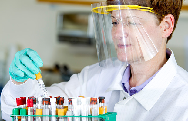
Medical Technologist Shelley selects a sample to be tested.
-
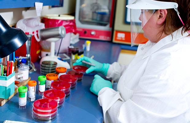
Becky prepares specimen culture plates in the microbiology department.
-
Cytotechnologist Sara processes Pap Smear specimens into slides to be analyzed using the BD Focal Point ™ GS Imaging System.
-
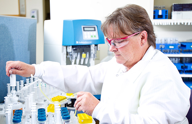
Melissa loads patient samples on an automated chemistry analyzer.
-
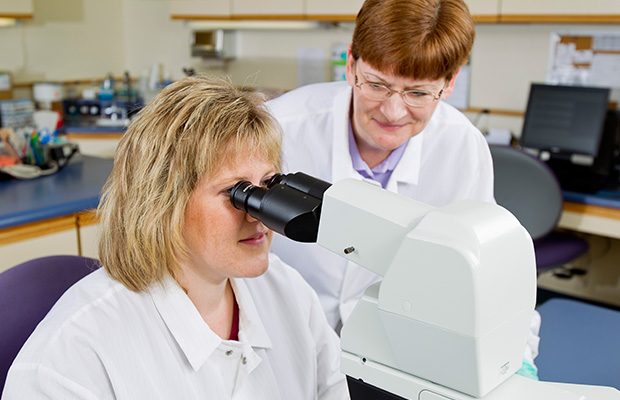
Jill and Shelley perform a manual differential cell count.
-
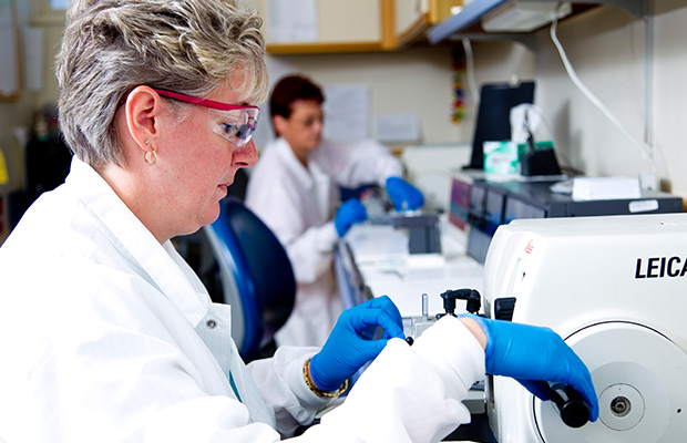
Joey prepares tissue specimens into slides to be interpreted by a pathologist.
-
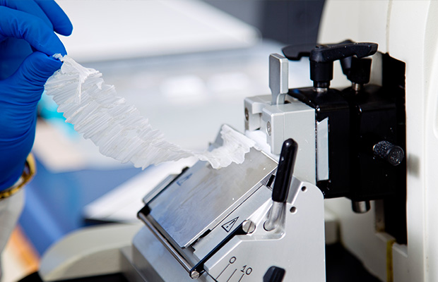
Histology technicians cut a surgical tissue specimen to make the slide.
-
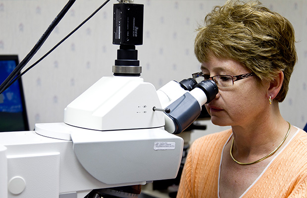
Julie C. Netser, M.D. Reviews a surgical case using the Ventana VIAS Image Analysis System.
-
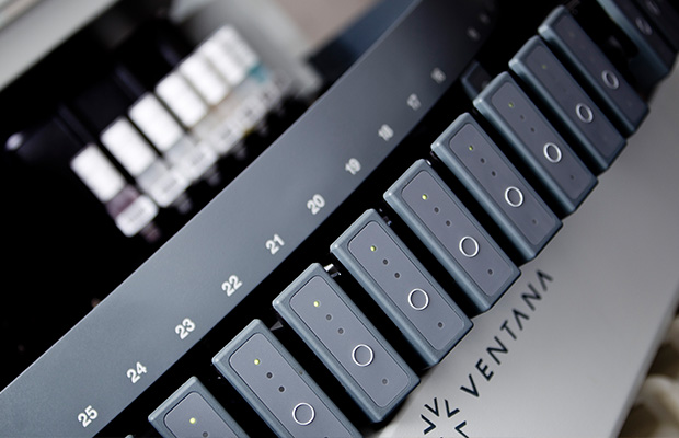
The Ventana Benchmark ULTRA staining platform aids pathologist in cancer detection.
-
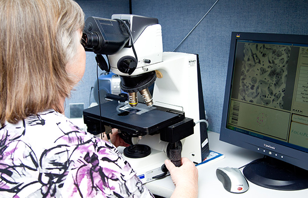
Cytotechnologist Sherie screens a Pap Smear using the BD GS Imaging System ™.


